Dislocation of the Elbow (Including fracture dislocation)
Simple elbow dislocations without associated fractures are generally stable despite complete rupture of all capsulo-ligamentous structures.
Conversely if associated with bony injuries treatment is more complex and prognosis worse.
Acute Simple Elbow Dislocations
Evaluate stability following reduction.
Gently move elbow through its range of motion. If the elbow appears to subluxate or dislocate, put in a backslab with elbow flexed 90° and do check x- ray (AP / Lat).
If elbow congruent in sling or backslab review 5-7 days AND re Xray!!!
If patients complains of any new symptoms re Xray!!!
If the elbow subluxates or dislocates in extension or is noncongruent on the radiographs made at the follow-up visit, pronate the forearm and reassess (pronation tends to increase the tension on the lateral soft-tissue constraints).
If stability is restored, a hinged brace or cast-brace is applied with the forearm in full pronation.
An extension block of 30° is sometimes necessary.
If the elbow requires an extension block of more than 30 to 45°, surgical repair should be considered.
Extension blocks should be gradually eased so that by three weeks the brace allows full motion. At each follow-up examination, re-evaluate in exactly the same manner.
Assessing stability
May need GA. Place arm in overhead position, the arm then resembles a leg, and the elbow resembles a knee.

Examine for:
-
Valgus - Perform with the forearm fully pronated so that posterolateral rotatory instability is not mistaken for valgus instability. This happens because the ulna and the radius rotate as a unit away from the humerus in response to valgus stress when the lateral collateral ligament is disrupted. Forced pronation prevents posterolateral rotatory instability because the intact medial soft tissues are used as a hinge or fulcrum, just as the periosteum is used for this purpose during the reduction of a supracondylar fracture in a child.
- Varus - Varus stress-testing is easiest to perform with the shoulder fully internally rotated
Both valgus and varus stress-testing are performed in full extension and 30° of flexion. Flexion unlocks the olecranon from the olecranon fossa
- Posterolateral rotatory instability - Posterolateral rotatory instability is diagnosed with use of the lateral pivot-shift manoeuvre of the elbow. If there is severe soft-tissue disruption, this test can have a false-negative result. With severe soft-tissue disruption, the elbow can sometimes remain dislocated even when it is flexed past 90°. If suspected, this problem can be avoided by the examiner using his or her thumb to prevent the elbow from fully dislocating (or limiting the degree of subluxation during the pivot-shift test).
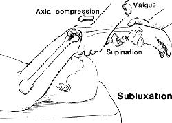
Lateral Pivot-Shift Manoeuvre
Currently, the most common method of performing this test is with the patient placed supine on the examining table with the affected extremity overhead. The elbow is supinated, and a mild-to-moderate forced valgus stress is applied while the elbow is flexed past approximately 40°. This manoeuvre results in apprehension or frank subluxation of the radius and the ulna in a rotatory fashion from the humerus. A visible, palpable clunk or an actual pivot shift may be elicited by this manoeuvre. If instability is not elicited, the test is still positive if there is apprehension, and the diagnosis can be made on this basis. The posterolateral rotatory drawer test involves placing the elbow in approximately 40° of flexion and applying an anterior-posterior force on the ulna and the radius to subluxate the forearm away from the humerus on the lateral side (pivoting on the intact medial ligaments).
Stress radiographs
AP plane with valgus and varus stress and the arm overhead as described above.
When valgus stress-testing is performed, the forearm must be held fully pronated with moderated force to prevent posterolateral rotatory subluxation and false-positive valgus instability.
With the shoulder in 90° of abduction and full external rotation, stress radiographs are also made with the arm in supination and pronation to detect posterolateral rotatory instability and to determine if the medial side opens up with pronation (indicating disruption of the medial soft tissues).
Elbow Facture-Dislocations
The Terrible triad
- Elbow dislocation
- Fracture of the radial head
- Fracture coronoid process
The fundamental goal in the management of fracture-dislocation of the elbow (a so-called complex dislocation) is the restoration of the osseous-articular restraints, thereby converting the injury to a so-called simple dislocation, which has been demonstrated to have a generally favourable long-term prognosis. In approaching the management of the terrible-triad injury, one must consider the individual injuries to the coronoid process, radial head, and collateral ligaments.
The coronoid process (see Coronoid process)
The coronoid process is a primary stabilizer of the elbow and is critical in the presence of a radial head fracture and ligament injury. Persistent instability is more likely as more of coronoid involved.
Exposure of the coronoid process depend on the associated lesions.
- Universal posterior skin incision, just lateral to the tip of the olecranon, permits deep access to both the medial and the lateral side of the elbow.
- An exception is a medial coronoid fragment (often part of a comminuted type-II or III coronoid fracture), which is more appropriately approached medially.
- Internal fixation of smaller (type-I or II) fragments may present not only technical problems but also problems that may jeopardize the tenuous blood supply to the coronoid process. An option is passing two braided sutures over the top of the small coronoid fragment, pulled out through drill-holes in the ulna, and tied over the bone. If the capsule is attached to the fragment, the sutures should be passed through the capsule as well.
- Larger (type-III and some type-II) fractures of the coronoid process require anatomical reduction and stable internal fixation.
Radial head fractures (see Radial head fractures)
Treatment
A concomitant dislocation of the elbow should be reduced, and the radial head fracture should then be managed on its merits.
Decision-making is influenced by:
-
Patient-related factors (age, bone quality, and activity level)
-
Fracture-related factors (fracture size, displacement, and location).
For example, an older patient with osteoporosis and a comminuted radial head fracture is a poor candidate for internal fixation, with radial head arthroplasty being the preferred option in the setting of an associated elbow dislocation.
Open reduction and internal fixation of
the radial head fracture should be attempted when it is
technically possible.
Acute excision of the radial head without replacement is
contraindicated when there is concomitant disruption of the medial
collateral ligament or the interosseous membrane. Displaced
fractures that block the rotation of the forearm and are
too small, comminuted, or osteoporotic for stable
internal fixation should be managed with fragment
excision. The two prerequisites for fragment excision
are evidence that the fragments to be excised do not
articulate with the lesser sigmoid notch of the ulna and
involvement of less than one-third of the radial head.
Delayed excision of the radial head can be used for an
isolated, comminuted, displaced fracture that involves
more than one-third of the radial head and does not
block forearm rotation, but it is not an option in a patient who
has had a fracture-dislocation. Arthroplasty of the radial
head is indicated for displaced, comminuted radial head
fractures when stable internal fixation is not
possible and the fracture involves more than one-third
of the radial head.
Operative Techniques
As in the case of a coronoid fracture, a posterior elbow incision is used. This approach decreases the risk of a cutaneous nerve injury compared with that associated with a separate medial incision. Disruption of the lateral collateral ligament complex and the common extensor muscles from the lateral epicondyle is commonly noted in patients with an elbow dislocation. This approach also simplifies the surgical exposure of the radial head. The interval between the anconeus and the extensor carpi ulnaris (the Kocher interval) is identified and developed. The lateral collateral ligament is incised at the midportion of the radial head, with the surgeon staying anterior to the lateral ulnar collateral ligament. The annular ligament often must be divided to improve exposure.
Internal fixation is performed with use of 1.5, 2.0, or 2.7-millimeter screws and plates or 3.0-millimeter cannulated screws, depending on the size of the fragment. If plate fixation is used, the plate should be placed on the nonarticular portion of the radial head, which is the lateral part of the radial head when the arm is in neutral rotation. Screws should be countersunk to avoid impingement with the lesser sigmoid notch. Threaded Kirschner wires may be useful for small fragments not amenable to screw fixation. Smooth Kirschner wires should be avoided due to their tendency to migrate during the postoperative period.
A radial head with a fracture that is not amenable to fixation should be replaced with a metallic radial head when there is a medial collateral or interosseous ligament injury. Silicone implants have been employed in the past; however, they have a high rate of failure due to fracture and fragmentation.
A word of caution: the coronoid fragment should not be removed when the radial head is excised.
Following fragment excision, open reduction and internal fixation, or radial head replacement, the lateral collateral ligament complex and the common extensor muscle origins should be carefully repaired back to the lateral epicondyle with use of heavy sutures through drill-holes or suture anchors. The fascial interval between the anconeus and the extensor carpi radialis also should be closed to augment lateral stability of the elbow. The elbow should be carefully evaluated for stability, and concomitant injuries (for example, those involving the medial collateral ligament or the coronoid process) should be repaired when appropriate.
Postoperative Management
Indomethacin (75mg daily (25mg TDS)) has been recommended for patients with complex elbow dislocations, to control postoperative pain and to potentially reduce the prevalence of heterotopic ossification. Avoid in patients with a history of peptic ulcer disease or a known allergy. An early range of motion within a safe arc should be initiated, depending on associated fractures and ligamentous injuries. Extension-splinting is initiated as soon as stability improves, and the splint is worn at night for twelve weeks.
Complications
Avascular necrosis might be expected following open reduction and internal fixation of most radial head fractures, as the fragments typically have a precarious blood supply or no blood supply. Fortunately, the fragments usually heal and late collapse is uncommon.
Nonunion is usually associated with avascular necrosis and seems to be more common in patients with a fracture involving the radial neck.
Malunion is usually a consequence of inadequate fracture fixation or collapse due to avascular necrosis. Osteoarthritis is seen as a consequence of articular cartilage injury from the initial dislocation, late instability, or articular incongruity.
Stiffness is a common sequela of a radial head fracture and may be due to capsular contracture or heterotopic ossification.
Late axial or valgus instability is uncommon unless the radial head has been excised.
References
JBJS-A 80:566-80 (1998)- Current Concepts Review - Fracture-Dislocation of the Elbow
JBJS - A 82:724 (2000) - Instructional Course Lecture - The Unstable Elbow
JBJS - A 84:547-551 (2002) - Posterior Dislocation of the Elbow with Fractures of the Radial Head and Coronoid