5th Metatarsal fracture - base
Jones fractures
Jones originally described an avulsion fracture from the base of the 5th metatarsal due to a forced inversion of the mid-foot. Later studies have demonstrated that it is rather a combination vertical and mediolateral forces focused over the 5th metatarsal base, not inversion (Kavanaugh 1978).
Often called a ‘tennis fracture’ the fracture occurs at this level due to the pull of peroneus brevis on the base of the 5th metatarsal. They are divided into three zones :
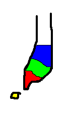 |
Type I - Avulsion fracture Type II - Jones fracture Type III - Stress fracture |
Type I (avulsion fractures) - are often small and are the commonest type of base of 5th metatarsal fracture. If minimally displaced these may be treated conservatively with a weight bearing plaster. Patients should be followed up as there is in an incidence of symptomatic non-union when screw fixation or excision of a small fragment should be undertaken.
Type II (Jones Fracture) - Although all base of 5th metatarsals are now termed jones fractures, Jones originally described the type II fracture. Due to the pull on the proximal pole and relative mobility of the joint a low threshold for acute fixation Jones fractures is appropriate.
An axial percutaneous cannulated screw is often the best method of fixation
Type III (Stress Fracture) - Metaphyseal fractures are most often stress fractures and should be treated as other stress fractures with relative rest and activity modification. Although in some circumstances operative fixation is appropriate caution is advised.
Os-peroneum is an ossicle seen in up to 15% of patients, and is normal.
The Jones fracture (Type II) is associated with a 66% delayed union rate (Kavanaugh 1978), and for this reason operative fixation is often advocated. Arntz et al reported a series of 40 operatively treated Jones type fractures with 36 achieving a good or excellent result at 3 years follow up.
Paediatric 5th Metatarsal Apophysis
In the paediatric skeleton an accessory proximal growth plate is seen in approximately 14% of patients.
This can easily be confused for a fracture.
In a similar manner to the accessory navicular (or os naviculari) patients may present following minor trauma with a tender 5th metatarsal, and the incidental finding of an accessory growth plate at the base of the 5th metatarsal.
The accessory epiphysis is seen most commonly between the ages of 11 and 14.
There are a number of types described by Rogers et al and they may have the appearance of a fracture :
Clinical examination and a careful history should distinguish this from a fracture.
If there is any doubt an MRI scan may be used to differentiate the two.
References
The Styloid Epiphysis of the fifth Metatarsal Bone; ROGERS JBJS(Am) 10 (2): 197. (1928)
The Jones fracture revisited Kavanaugh-JH; Brower-TD; Mann-RV JBJS(Am) 1978 Sep; 60(6): 776-82
Fractures and fracture-dislocations of the tarsometatarsal joint; Arntz-CT; Veith-RG; Hansen-ST JBJS(Am) 1988 Feb; 70(2): 173-81