Tibial Plateau fractures
Usually high energy injuries caused by varus or valgus stress +- axial loading. Important to note integrity of soft tissue envelope and exclude associated neurovascular injuries in higher grade fractures.
Classification
 |
I - Split fracture of the
lateral tibial plateau without articular depression II - Split depressed fracture of the lateral tibial plateau III - Isolated depression of the lateral plateau IV - Fracture of the medial plateau V - Bicondylar plateau fracture with varying degrees of articular depression and displacement of the condyles VI - Bicondylar tibial plateau fracture with diaphyseal metaphyseal dissociation |
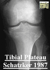
Radiology
AP Lateral of knee usually sufficient for diagnosis.
40° internal and external oblique views - can be used if a fracture is suspected but not seen on above.The internal oblique shows lateral plateau, external oblique shows medial condyle and plateau.
CT Scan or MRI - If considering surgery to evaluate anatomy of fracture lines. +- ligamentous injury
Angiography - Have low threshold to perform angiography. High-energy injuries, fracture–dislocation patterns, unexplained compartment syndromes, and Schatzker 4, 5, and 6 fractures should lower the threshold for obtaining an arteriogram.
Management
Initial management is directed to soft tissue, associated injuries and actively excluding neurovascular injury. Observe closely higher grade injuries for development of compartment syndrome.
Several factors affect choice of treatment: Aim to achieve stable congruent joint, avoid infection/ skin problems
| I | If displaced - 1.open reduction and internal fixation or 2. Arthroscopy and if meniscus intact, closed reduction and percutaneous fixation. Need to be sure meniscus not interposed in fracture. |
| II | Elevation depressed fragment +- bone grafting and buttress plate fixation of condylar split. +- arthroscopic assistance ( beware fluid and compartment syndrome) |
| III | If the area of articular depression is small and the joint remains stable, treatment is nonoperative. If the joint is unstable in a physiologically young patient, surgical treatment is usually indicated. Elevation of depressed fragments +- arthroscopic assement joint. Followed by bone grafting and percutaneous screw fixation to support joint and graft. |
| IV | Usually high energy. It is often associated with knee dislocations and neurovascular injuries. Asses soft tissue injury carefully (MRI +- arteriography). Nonoperative only for nondisplaced fractures. Risk varus malunion. Surgery - closed reduction and percutaneous screw fixation. Unstable injuries may require fixation through a midline or medial patellar approach and exposure of the large medial fragment. simple lag screws may not be sufficient fixation and may require a buttress plate If the intercondylar eminence with the attached cruciate ligament is avulsed, it should be reduced and fixed |
| V & VI | Very diverse group of injuries. Aim to achieve stable, aligned, mobile, and painless joint and to minimize the risk of post-traumatic osteoarthritis. Status of soft tissues may dictate management options. Options - 1.Conservative, maintain alignment avoids risk of infection and function may be surprising despite X-ray appearances 2. Open reduction and internal fixation, risk infection and wound problem. Double buttress plating mechanically is the strongest but increases soft tissue morbidity and compromises fracture vascularity 3. External fixation, Hybrid or ring fixator preserve soft tissue envelope, preserving fracture vascularity, risking displacement and pin site sepsis. |