Paediatric ankle Overview
The Physis is a relatively weak point compared to adjacent bone, ligaments or tendons. This accounts for markedly different injury patterns in the growing child compared with those seen in adults.
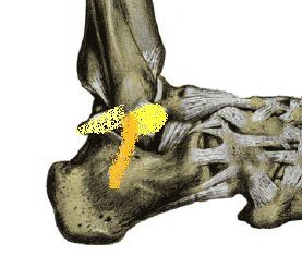
Acute traumatic events often result in fracture at the physis, or apophyseal avulsion, in a child, leaving the ligaments intact.
Anatomy
Anatomically, the ankle joint is best thought of as a hinge, with the talus mortised between two pillars.
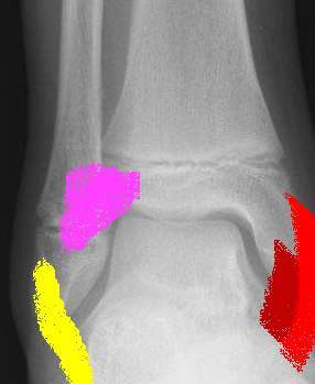 |
Each pillar has a bony and ligamentous component
Immediately above the joint, but below the tibial physes, the fibula is attached to the tibia by the syndesmotic ligaments.
Medially, the deltoid ligament is composed of a deep and superficial layer.
Note the medial and lateral ligaments arise directly from the epiphyses. |
 |
The lateral collateral ligament consists of three ligaments
|
Radiographs
AP - Antero-posterior
Lateral
Mortise view - taken with 15-20° internal rotation of leg
Comparison views of the unaffected leg may be necessary because the numerous ossification centres in the foot and ankle may make interpretation difficult.
 |
 |
|
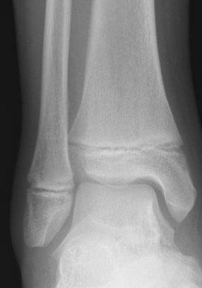
12YO Male AP Ankle |
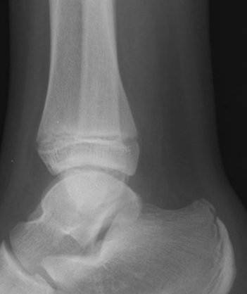
12YO Male Lat Ankle |
References