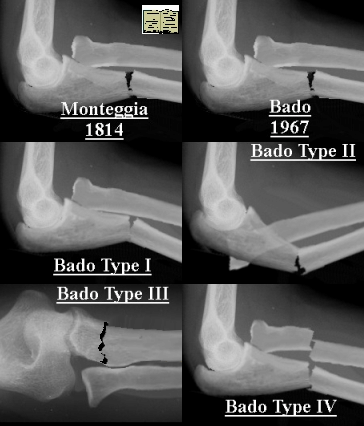
The Monteggia fracture
Classification
Monteggia (1814) fractures are classically described as a dislocation of the radial head and fracture of the ulna. Bado subclassified this into four types.
 |
Bado ClassificationI -Anterior dislocation of the radial head and fracture of the diaphysis of the ulna at any level, with anterior angulation of the fracture fragments. II - Posterior or posterolateral dislocation of the radial head and fracture of the diaphysis of the ulna, with posterior angulation of the fracture fragments. III - Lateral or anterolateral dislocation of the radial head and fracture of the metaphysis of the ulna. IV - Anterior dislocation of the radial head and fracture of the proximal third of the radius and ulna at the same level. |
Treatment Principles
The generally accepted treatment of the Monteggia fracture in adults is internal fixation of the ulna.
Treatment in children depends on the character of the ulnar fracture:
Plastic deformation - requires reduction of the ulnar bow under GA to
achieve stable reduction of the radio-ulnar joint.
Incomplete (greenstick or buckle) - require closed reduction and
casting. Nearly all Monteggia fractures in children (Bado types I and III) are
most stable when immobilised in 100 to 110° of flexion and full supination.
Nearly complete greenstick or one associated with a radial fracture (Bado type IV) - Consider operative fixation with an intramedullary K wire
Complete transverse or short oblique - Are often in bayonet apposition, with malalignment or shortening. Reduction may be difficult and is usually unstable. K wires are useful for obtaining reduction by manipulation of the proximal fragment, and for holding the reduction.
Long oblique or comminuted - May develop shortening and malalignment even when fixed with intramedullary wires, and therefore recommend the use of a short plate and screw.
Type I (70%)
Anterior dislocation of the radial head with an associated ulnar diaphyseal
fracture, usually a short oblique or a complete greenstick pattern.
Three major mechanisms of injury proposed:
1. Direct blow posteriorly breaks the ulnar diaphysis and then forces the radial head into an anterior dislocation. no evidence to support this injury mechanism in children.
2. Hyperpronation mechanism - hyperpronation force applied to the outstretched arm fractures the ulnar shaft and forces the radial head to dislocate.
3. Hyperextension mechanism - Most currently accepted
one. Injury occurs in three phases. First, elbow hyperextension occurs as the
child tries to arrest a fall on an outstretched arm. Secondly, during elbow
hyperextension, biceps contraction resists the extension moment and dislocates
the radial head. Lastly, after radial head dislocation, the body weight is
transmitted to the forearm, concentrated on the ulnar diaphysis that fails in
tension, producing a complete oblique or a greenstick fracture.
Treatment
Usually nonsurgical in children and follows three steps. Ulnar diaphyseal fracture anatomic reduction is the key to success. Once the ulnar fracture is reduced to length, the radial head easily reduces. Elbow flexion of 110–120° diminishes biceps force on the proximal radius. Moulded above elbow cast 3 weeks followed by below elbow 3 weeks. Check x-ray 1 week to ensure reduction maintained. Surgery indicated if unable to anatomically reduce or unable to hold ulna reduced out to length see above principles.
Type II (6%)
Apex-posterior ulnar angulation at the olecranon-diaphyseal junction, with posterior radial head dislocation.
Mechanism of injury longitudinal, proximally directed force up the forearm with the elbow
semi-flexed causes the posterior ulnar cortex to fail.
Treatment
Closed reduction is done by longitudinal traction with the elbow extended.
Direct anterior directed pressure on the radial head to reduce.
Since this is a flexion injury with the ulnar anterior cortex usually intact,
the ulna is most stable with the elbow in extension. Moulded cast with elbow in
extension to maintain ulna. Because the ulnar fracture
is usually metaphyseal, healing is rapid and 3 weeks in a cast should be
sufficient. Advise parents that it may take some time for elbow flexion to
return. Operative indications similar to type I.
Type III (23%)
Type III is the one most commonly associated with irreducibility of the radial head
because of interposition of the annular ligament. The incidence of posterior interosseous nerve injury is high with this lesion.
The nerve deficit usually completely resolves rapidly and spontaneously.
Mechanism of injury - varus stress on an extended elbow.
Ulnar failure as a greenstick olecranon fracture occurs first. The radial head
then dislocates laterally or anterolaterally.
Treatment
Reduction involves reversing the injury mechanism. The elbow is hyperextended to stabilize the olecranon. With the elbow in extension, a valgus force is applied to the olecranon to correct (or slightly overcorrect) the greenstick fracture. The radial head will often reduce spontaneously. Local pressure directed medially over the radial head may be necessary to complete the reduction.
Immobilize
similar to Type I. Some clinicians recommend that, because of the tendency for the olecranon to re-deform, the
arm should be immobilized with the elbow in extension in a long arm cast with a
valgus moment applied to the ulna. Four weeks is usually enough to
heal the greenstick injury.
Operative indications similar to type I.
Type IV (1%)
Fracture
radial and ulnar shaft with dislocation of radial head. Mechanism of injury probably similar to type
I.
Treatment
Usually requires surgical stabilization of the radius and ulna fractures. In younger children, intramedullary wires are usually sufficient to stabilize the forearm fractures. Plate fixation of the fractures may be required in older children. Immobilize above elbow 3 weeks, then below elbow 3-4 weeks.
Delayed presentation/ missed fractures:
Patients chosen for treatment should be less than 12 years of age and should have absence of radial head changes. Surgery is indicated for patients who have the following:
1. progressive radio-capitellar subluxation or dislocation
2. progressive elbow valgus deformity
3. limited range or forearm motion
4. pain at the malaligned radio-capitellar or radio-ulnar articulations.
Complications
Complications are quite uncommon.
Motion usually returns fully within 3 months in promptly treated cases.
Nerve injuries are rare, most common in Type III lesions with posterior interosseous nerve involvement. Spontaneous resolution of this neuropraxia is the rule by 9–12 weeks post injury.
Tardy ulnar palsy is a rare neuropraxia that occasionally is associated with an elbow valgus deformity seen with a long-standing unreduced radial head dislocation. Injuries to the median nerve are quite rare.
Periarticular ossification may uncommonly be seen in cases of chronic dislocated radial heads. The ossification tends to resorb post reduction. Myossitis ossificans can accompany the lesion if a radial neck fracture is also present.
References
Wilkins, Kaye E. D.V.M., M.D.. Changes in the Management of Monteggia Fractures. Journal of Pediatric Orthopedics. 22(4):548-554, July/August 2002.
OPERATIVE FIXATION OF MONTEGGIA FRACTURES IN CHILDREN -David Ring; Peter M.
Waters; JBJS - 78 B: (734-739) 1996